Visual systems beyond a single visual network AI
The visual system is a collection of interacting regions with different spatial organizations and response properties. A broad “Visual” label in a functional atlas compresses that diversity. It is useful for identifying a large-scale footprint, but is too coarse to identify a particular visual computation or stimulus preference.
Visual space is mapped repeatedly
Early visual cortex includes retinotopic maps: neighboring cortical locations represent related positions in the visual field. V1, V2 and V3 are not interchangeable parcels, and visual field maps extend into lateral, ventral, dorsal and parietal cortex. Wandell, Dumoulin and Brewer describe their organization and measurement.
Retinotopy is established using stimuli that systematically sample visual position. A parcel’s correlation with other visual parcels during rest does not identify its precise field map, receptive field or category preference.
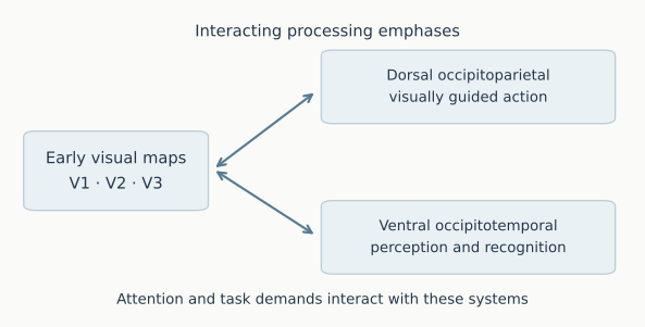
A conceptual organization, not an anatomical tract diagram or an atlas boundary map. Visual processing includes extensive recurrent interactions.
Processing streams and category preferences add other distinctions
The distinction between ventral pathways supporting perception and recognition and dorsal pathways supporting visually guided action is an influential framework (Goodale and Milner). These are broad functional emphases, not two isolated channels or an exhaustive account of vision. Attention, eye movements and task demands also recruit systems beyond a coarse visual-atlas label.
Category localizers ask a different question: which regions respond preferentially to one class of images relative to a specified comparison? Face-selective fusiform responses provide a classic example (Kanwisher and colleagues). Preference depends on the contrast and should not be equated with an exclusive role in one category.
Match the anatomical claim to the measurement
The Studyforrest localizer supplies controlled image categories and fixation periods. Pooling visual blocks emphasizes processes shared across stimulation; category contrasts test finer differences. A cortical ROI average can dilute a response confined to a smaller selective region, so a weak network-level category difference need not imply absent category selectivity at voxel level.
For a learned network pole, a strong match to the broad visual reference supports a visual-oriented spatial description. It does not by itself distinguish early retinotopic processing, face recognition, scene processing or visuomotor coordination. Those claims require finer anatomical mapping, relevant experimental contrasts and checks on spatial resolution and signal coverage.