Registration, resampling, and anatomical coverage AI
Extracting an ROI time series assumes that the atlas region and the functional measurements refer to the same anatomy. Registration, resampling and coverage checks address different parts of that assumption. Passing one does not establish the others.
Alignment estimates a correspondence
Registration estimates a transformation between images. A rigid transformation changes position and orientation; an affine transformation can also change scale and shear. Nonlinear registration permits spatially varying deformation. These choices describe different capacities to match anatomy, rather than interchangeable levels of image resolution.
For fMRI, a common route aligns functional images with a participant’s structural scan, then maps that scan to a template. Transformation conventions and composition order matter. The FSL FLIRT documentation describes affine registration and its application.
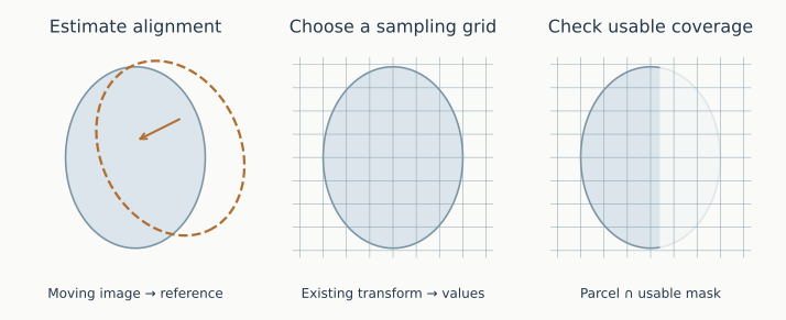
Schematic anatomy and grids. A matching grid alone cannot establish correct anatomical alignment.
Resampling evaluates the mapped image
Resampling assigns values to a chosen target grid using an existing spatial mapping. A voxel value is estimated at each target location; this does not itself estimate a new anatomical correspondence. For example, Nilearn’s resample_to_img explicitly assumes that registration has already been established.
Interpolation must respect the data. Discrete atlas labels usually require nearest-neighbor interpolation so that label numbers are not blended. Continuous intensity images use different interpolation rules. Changing the grid can alter parcel voxel counts and boundary sampling without improving the underlying alignment.
Coverage limits the measurement
A parcel may extend outside the usable functional mask or contain substantial signal dropout. Coverage can be summarized as the proportion of its voxels inside that mask, with the mask definition and threshold recorded. An average from a small surviving portion may represent a different sample of tissue from another participant’s parcel average.
Coverage is therefore a measurement check, not activation confidence. High coverage does not prove alignment or signal quality, and low coverage does not establish neural abnormality. Inspect anatomical overlays alongside the mask and signal traces before attributing unusual network weights to functional differences.